Doctors may perform a complete physical examination and medical history review to diagnose symptoms of sinus infections. Other scientific diagnostic methods may also be used. For instance, x-ray, computerized tomography (CT), ultrasound and cultures.
X-ray uses invisible electromagnetic energy beams to capture images of internal bones, organs and tissues. Quality and accuracy of x-ray depends on doctors’ ability and technique to read the images correctly. In many cases, x-ray of sinuses does not capture symptoms of sinus infections.
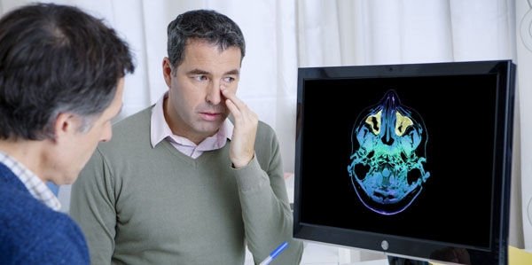
CAT or CT scan, a noninvasive reliable medical test,uses computer technology and X-rays to generate multiple vertical and horizontal slices (cross-sectional images) of affected body part. It generates real time pictures. These scans reveal more details of the body as compared to the X-rays. The facial CT scan produces pictures of paranasal sinus cavities. Sinus CT is performed to identify inflammatory diseases, gather anatomical information for sinus surgery to remove tumors in sinuses or nasal cavities, and identify sinuses filled with fluid and thick sinus membranes. CT is an easily available service requiring lesser time than magnetic resonance imaging (MRI). This cost-effective, accurate and painless method can capture pictures of blood vessels, soft tissues and bones simultaneously. Quick and simple CT scans are a good choice for emergency cases.
Ultrasound, a noninvasive tool, is also used to identify symptoms of sinus infections. The tool is reliable, quick and less costly than a CT scan. However, results of ultrasound are not as good as that of the CT scan. Ultrasound is generally not used for sinusitis diagnosis.
Cultures of sinus or nose fluid are prepared in the laboratory to help in diagnosis.
Doctors may also conduct blood tests.
Physicians may recommend a visit to an ear, nose and throat (ENT) specialist if sinusitis recurrent and severe. The specialist may conduct more tests. ENT specialists use nasopharyngoscope, a flexible fiberoptic tube, to examine the passage into the sinuses (OMC) and the nasal passages. The tube inserted in the nose helps in visualizing structural deformities like tonsils, enlarged adenoids, nasal polyps and deviated nasal septum. The specialist may drain the sinus to examine the organisms. Sinus draining is an invasive procedure. It requires a needle, which is inserted into the sinus through gum or skin to extract the fluid. The fluid is sent to the laboratory for culture preparation and identify bacteria. However, it is uncommon because of high level of discomfort.